For my 4th week of observations I spent the majority of my time photographing some of the organisms that I have already been posting about. I took nearly 80 photographs of various lifeforms, some new to me and some not. I have posted some of the best photos below.
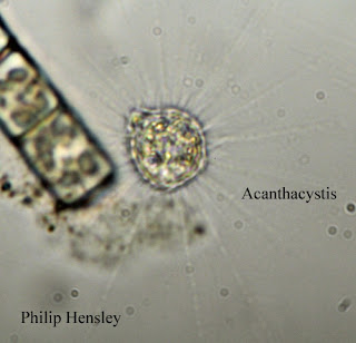
This photo shows an Acanthacystis (Patterson, 2003).
There were many of these found all over the tank.
Another Acanthacystis found floating just above the
remnants of the beta food pellet (Patterson, 2003).
Each of the images of Acanthacystis clearly show the
various vacuoles and chloroplasts inside them(Patterson,
2003). I expected to see something going in or coming
out of this one because of the protrusion in the upper left,
but nothing occurred.
This image shows a Chroococcus (Patterson, 2003).
Note the bright green chloroplasts, showing this to be
an autotroph.
In addition to these organisms, I also saw several Rotifers. These were later identified to be Euchlanis sp. (Patterson, 2003). They were fast moving, and very agile. These pictures were taken of the same organism. You can clearly see the scenery changing around it as it quickly made its way across the Microaquarium.
This photo was taken when I first noticed the Rotifer swimming
across the tank. I zoomed in to get a closer look. Even at this
magnification it is easy to see some of the internal structures
of Euchlanis sp. (Patterson, 2003).
Here the organism is investigating some residue at the
bottom of the Microaquarium.
This image clearly shows the forked tail of the Euchlanis
sp. The tail was rarely spread out, but it resembled
a snake's tongue whenever it was opened.
I will obtain a few more edited images to post to the blog next week. As I said before, I have roughly 80 total images and need to narrow down the remaining ones to share you everyone.


great photos!
ReplyDelete