This week's Microaquarium observation yielded several sightings of a previously unseen (at least to me) work-like organism. It was large in diameter, and also very long when compared to other organisms in the tank. The body was encircled by several groupings of fine "hairs," occurring in clusters of 4 that encircled each seam or joint of its body. At the anterior end it had 2 very long flagella that constantly flapped forward and backward. Its head or mouth part was nearly tapered to a point. It was actually plainly visible to the naked eye. The worms were spread along the bottom of the tank. Most appeared to be near the surface of the sediment layer that had settled at the bottom of the Microaquarium. I observed 3 of them that were clumped closely together for roughly 45 minutes. The largest one was the least active, but I was able to watch its transparent organs at work. Its intestines pulsated rhythmically, and I was actually able to watch other dead organisms passing through the various parts of its body, and even saw them evacuated from the worm. My inner nerd was totally mesmerized...VERY COOL!
Also making the "Recently Sighted" list this week are several Desmids. I had commented earlier in the blog about noticing some "green bananas" in the Microaquarium, but this time I spotted 10 to 12 lager desmids. They were still green in color, but were much longer than the "bananas," and were straight, not bent in the center. They moved very slowly, and appeared to have the same flagella at each tip like the others had.
Interestingly enough, I only noticed 1 or 2 rotifers this week. Not sure if the were between generations or nt, but these had been the most abundant organism for the past 2-3 weeks, and now I couldn't find them no matter how much I looked. However, the number of Bodos in my tank had increased. They were in almost every frame I had searched in, mostly in the upper region of the tank, not near the sediment surface. Perhaps they were hunted at that level, or they may just prefer the upper region.
I tried to capture an image of a Dinoflagellate, but was unable to get a focused picture due to its constant movement. I could clearly make out the groove around it's midsection from the flagella. It swam in a side to side motion, and moved rather quickly, as I could not get the picture to develop. Still neat to see.
I believe this was the final opportunity to view the Microaquariums this term. I was told that these organisms will be added to a small pool maintained by the Biology department behind the Hessler Building. It's time to see if thee microorganisms can truly adapt to some new surroundings. GOOD LUCK, little guys!!
Sunday, November 17, 2013
Bibliography Page:
Bibliography:
Rainis K, Russell B. 1996. Guide to Microlife. New York: Franklin Watts Publishing. 287p.
Pennak, R. 1989. 3rd Edition. Freshwater Invertebrates of the United States: Protozoa to Mollusca. New York: John Wiley & Sons. 628p.
Patterson, DJ. 2003. Free-Living Freshwater Protozoa: A Color Guide. Washington D.C.: ASM Press. 223p.
Saturday, November 9, 2013
Pictures of Third Creek Lifeforms
For my 4th week of observations I spent the majority of my time photographing some of the organisms that I have already been posting about. I took nearly 80 photographs of various lifeforms, some new to me and some not. I have posted some of the best photos below.
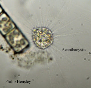
This photo shows an Acanthacystis (Patterson, 2003).
There were many of these found all over the tank.
Another Acanthacystis found floating just above the
remnants of the beta food pellet (Patterson, 2003).
Each of the images of Acanthacystis clearly show the
various vacuoles and chloroplasts inside them(Patterson,
2003). I expected to see something going in or coming
out of this one because of the protrusion in the upper left,
but nothing occurred.
This image shows a Chroococcus (Patterson, 2003).
Note the bright green chloroplasts, showing this to be
an autotroph.
In addition to these organisms, I also saw several Rotifers. These were later identified to be Euchlanis sp. (Patterson, 2003). They were fast moving, and very agile. These pictures were taken of the same organism. You can clearly see the scenery changing around it as it quickly made its way across the Microaquarium.
This photo was taken when I first noticed the Rotifer swimming
across the tank. I zoomed in to get a closer look. Even at this
magnification it is easy to see some of the internal structures
of Euchlanis sp. (Patterson, 2003).
Here the organism is investigating some residue at the
bottom of the Microaquarium.
This image clearly shows the forked tail of the Euchlanis
sp. The tail was rarely spread out, but it resembled
a snake's tongue whenever it was opened.
I will obtain a few more edited images to post to the blog next week. As I said before, I have roughly 80 total images and need to narrow down the remaining ones to share you everyone.
Sunday, November 3, 2013
3rd Observation
Last Wednesday, October 30th, I made my 3rd observation of the Microaquarium. The previous day our instructor had added a small food pellet to our tanks. I made the prediction that the amount of activity seen in the tank would increase dramatically. This theory proved to be correct. There were numerous lifeforms swimming all around the food pellet, and several new organisms that I had not noticed before. I spent approximately 2.5 hours in the lab studying many different varieties of life.
Among these organisms were many Rotifers. Euchlanis sp. was the most common form of these (Patterson 2003). I noticed for the first time that these Rotifers had tails that were forked. I previously thought that it was a single prehensile-type appendage, but was surprised to see that it was actually two individual structures. An even more abundant life form in my sample this week was a small organisms that was shaped like a sphere with many "spines" sticking out from the center. It looked much like a drawing of a sun might look. After several minutes of debate, and the help of D.J. Patterson's book, it was identified as being an Acathaystis. We took several detailed pictures of the organism and were able to narrow our options down. I will add the pictures we took throughout the next week, after they have been properly edited and re-sized.
I also was able to capture pictures of a Lacrymaria (Patterson 2003). It had the shape of an oval when it was stationary, but when it moved it could elongate its body, and appeared like a small squash might look. Its mouth parts and large vacuole became clearly visible when it elongated its body making it easier to identify. Again, I will add pictures in the coming week. A Bodo was also seen swimming around in the tank this week (Patterson 2003). It is a very small organism with two flagella, one much longer than the other. It contained a small vacuole. I only observed one of these, but I am certain there are more.
There were also several cyanobacteria sen in the Microaquarium this week. The most common one I noticed was Chroococcus (Patterson 2003). It took the shape of two green cells joined together with a thick membrane enclosing both of them. I did not notice any movement from this organism.
After observing these things, I uploaded my images to Photoshop, and began to narrow down which images I will be posting to this blog. I started with nearly 60 images, and though I have finished the final selection process, I must now go back and re-size and enhance the images for better quality and resolution. Check back by Thursday for the updated images of these and hopefully other organisms.
Sunday, October 27, 2013
2nd Observation: (Tuesday October 22, 2013)
Last Tuesday I made my second observation of my Microaquarium. I noted several organisms living in every region of the tank. The lifeforms had settled down a lot since the initial examination last week. Most of the activity was focused around the moss. I noticed several Euglenoids and Desmids in particular (Rainis & Russell, 1996). The Euglenoids were identified to be the Phacus species, and they movedquite rapidly using their flagella. They had a wide range of travel, moving up, down, left or right with relative ease and smooth motions. The Desmids were identified to be the species Chlorophyta. They moved slowly, with very subtle motions of the flagella on opposite ends of their banana-like shape. They were green, meaning they contained chloroplasts. There were also several other super small organisms that I was unable to identify. This week I will have the aid of the instructor to assess exactly what those were. They appeared to be single-celled lifeforms that simple shuttered or vibrated back and forth. Like shaky spheres.
Bibliography:
Rainis K, Russell B. 1996. Guide to Microlife. New York: Franklin Watts Publishing. 287p.
Pennak, R. 1989. 3rd Edition. Freshwater Invertebrates of the United States: Protozoa to Mollusca. New York: John Wiley & Sons. 628p.
Patterson, DJ. 2003. Free-Living Freshwater Protozoa: A Color Guide. Washington D.C.: ASM Press. 223p.
Sunday, October 20, 2013
Setup and 1st observation of Microaquarium
10/15/2013
Today I assembled my Microaquarium for Botany class. The purpose of the Microaquarium is to help our class learn about the wide range of lifeforms and diversity found in the smallest of places. We will be observing organisms on both a micro and macro scale.
The entire process of setting up the Microaquarium didn't take very long, about 15 minutes, once everyone knew where to find all of the materials they needed. I obtained a glass tank, a lid, and a tank stand and assembled them. Then, I was instructed to obtain 3 colored sticker dots, to place on the left margin of my glass tank. The first dot represented the lab section I was in, while the second dot represented which table I was sitting at, and the third dot represented my seat number at my table. I then wrote my initials on the dots to ensure I would be able to identify my Microaquarium in the future.
There were several containers laid around the lab with various numbers assigned to them. These were filled with the different water samples the instructor had collected. Unexpectedly, I got to choose which water source I wanted to use for my Microaquarium project. I had previously asked the instructor if there would be water samples made available from Third Creek, a notoriously polluted area located right next to the university's agricultural campus that I had always been curious to sample, and the instructor stated that he would get some samples if we really wanted to study them. Naturally, I selected the Third Creek sample to add to my Microaquarium. The information for the sample area was:
Third Creek in Tyson Park, Knox Co., Knoxville, TN
area in partial shade
N30 57 13.53 W83 56 32.37
824ft. on 10-14-2013
I next used pipettes to apply the sample into my Microaquarium tank, making sure to collect some sediment
material from the bottom of the container, and also collecting water from the surface to ensure I had an accurate representation of the various lifeforms that lived in the different strata of the water sample. Next, I added some samples of plant life. One form was Utricularia gibba, which is a carnivorous, flowering plant. It was originally from Spain Lake (N 35o55 12.35" W088o20' 47.00), Camp Bella Air Road, East of Sparta, TN in White County, and is also grown in water tanks outside Hesler Biology Building at The University of Tennessee and was collected on 10/13/2013. The other plant form was a moss, Fontinalis sp., collected from the Holston River along John Sevier Hwy under the I-40 Bridge. It came from an area with partial shade exposure (N36 00.527 W83 49.549) at an elevation of 823 feet, and it was collected on 10/13/2013.
Once the plants were properly positioned inside the tank, I was able to place the tank onto the microscope's
observation deck and study my sample for signs of life. Immediately, I noticed several things moving around in the sample. As I adjusted the focus of the microscope I began to see several multi-cellular organisms. I noticed at least two specimens that appeared to be some sort of mite. They seemed to wander all over the tank. I next noticed movement around the moss plant. I was not able to clearly identify what they were, but hope to be able to next week during my next observation. They were green-colored, and a couple appeared to be shaped like bananas. As I looked closer, I found many single-celled lifeforms inhabiting the upper area of the tank. They seemed to spin end over end, never really slowing down or stopping to clearly see what their structure truly was. I assume that the next viewing will show the organisms a bit more settled.
I plan to make my next observation on Tuesday, October 22nd, between 12-1pm.
Subscribe to:
Comments (Atom)


